HOW IS GLAUCOMA DIAGNOSED?
The diagnosis (or exclusion) of glaucoma requires a detailed, comprehensive examination of the eye. Your doctor will do the following examinations:
1.A routine vision test that requires reading letters from a chart
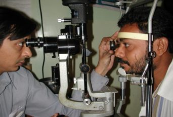
2.Slit lamp (microscope) examination
3.Measurement of the pressure in the eye usually using the applanation tonometer attached to the slit-lamp microscope. A hand held version of the same instrument is acceptable. It may be necessary to obtain multiple readings of the pressure during the course of the day and night.
4.Examination of the angle of the eye using a gonioscope. Steps 3 and 4 require the use of a drop to eliminate the sensation in the eye. The drop may burn a little bit.
Drops in the eye to dilate the pupil to facilitate:
5.Examination of the optic disc and the back of the eye (retina). Obtaining a stereoscopic view on the microscope using a hand held lens is the best method.
6.Newer computerized methods of examining the optic disc as shown above may be ordered. This is literally a computerized scan of the optic disc.
7.If glaucoma is suspected, then to confirm the diagnosis, the doctor will obtain an automated field test (perimetry test). Some patients may have difficulty doing this test for the first time. It may also be necessary to obtain several such tests as a baseline for future comparison. Considering the importance of the test ANY automated perimeter is NOT acceptable. The field test (perimetry) is a subjective test and it is important to have a calibrated machine with an appropriate normal database against which to compare your results.
In some cases a diagnosis may not be possible on one visit. In very early cases it may be necessary to repeat the entire examination after a period of observation.