
How Common is Orbital Cellulitis?
Patient Presentation
A 9-year-old female came to clinic with swelling of the left eye for 1 day and 4 days of fever. She had previously been evaluated in a walk-in medical care office and treated for conjunctivitis with antibiotic drops two days previously. She currently had photophobia, blurred vision, headache above both eyes, but no oral, facial or ear pain. She had had rhinorrhea for the past week and denied trauma or insect bites. The past medical history and family history were non-contributory. The review of systems was otherwise negative.
The pertinent physical exam showed a female with obvious facial swelling. Her vital signs were normal except for a temperature of 101.5° F. Her growth parameters were 10-25%. She could not open her left eye and the swelling and erythema extended above the eyebrow, into the cheek about 2 cm and laterally about 1 cm from the eye. With lid retraction, she had marked bulbar and palpebral conjunctival injection, photophobia, painful extraocular movements and proptosis. She had some limitations to inward gaze but it was difficult to assess because of pain. She was tender over the ethmoid sinuses with purulent rhinorrhea. She had no dental pain and the rest of her examination was normal. Thediagnosis of orbital cellulitis was made. The radiologic evaluation of computed tomography of the orbits showed a medial wall, retrobulbar abscess formation and ethmoid sinusitis. The patient’s clinical course showed that Ophthalmology was consulted who placed her on intravenous broad-spectrum antibiotics and eye rinses. A respiratory culture grew Haemophilus influenza type B and the patient was placed on Ciprofloxacin for a 2-3 week antibiotic course. Outpatient follow-up at 12 days showed no opthalmological deficits and the patient was otherwise doing well.
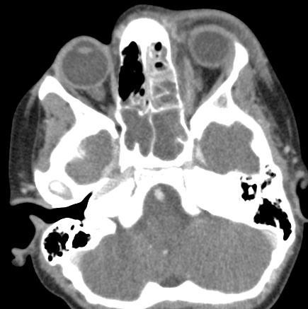
Figure 92 – Axial image from a CT scan of the orbits performed with intravenous contrast demonstrates sinusitis of the left ethmoid sinus and both sphenoid sinuses. There is left sided prespetal cellulitis anterior to the left orbit. Furthermore, there is a left sided subperiosteal abscess between the medial wall of the left orbit and the left medial rectus muscle.
Discussion
Orbital cellulitis is a serious infection whose complications can include meningitis, intracranial abscess, cavernous sinus thrombosis, carotid artery occlusion and vision loss. Orbital cellulitis itself is usually a complication of rhinosinusitis particularly of the ethmoid sinuses but also trauma.
Haemophilus influenza type B usually has been the prevalent causative organism withStaphlococcus aureus and viridins streptococcus also being common causes. The microbiology appears to be changing though. A 25-year study shows that although immunization against Haemophilus influenza type B and pneumococcus have decreased the cases of invasive infections such as meningitis, epiglottitis and bacteremia, immunization does not appear to decrease the cases of orbital cellulitis. Yet a recent 5 year review of patients at a children’s hospital found that Streptococcus anginosus was an emerging pathogen in pediatric orbital infections. See To Learn More below.
To learn more about the physical characteristics distinguishing periorbital (preseptal) cellulitis from orbital cellulitis click here.
Learning Point
Overall orbital cellulitis is generally an uncommon pediatric problem, especially in contrast to periorbital cellulitis. Exactly how uncommon depends on the research study but it is important to consider its possibility in any child presenting with swelling around the orbit. Some recent studies are abstracted below.
In a 10-year retrospective study of 52 patients less than 2 years of age with rhinosinusitis, orbital complications were evaluated and none had orbital cellulititis. This would be ~0 patients/year.
In another 10-year retrospective review, 6 patients with orbital complications secondary to acute sinusitis were evaluated and 1 had orbital cellulitis. This would be ~0.1 patients/year.
In a third 10-year retrospective review, 83 patients with preseptal or orbital cellulitis were evaluated and 14 had orbital cellulitis. This would be ~1.4 patients/year.
In a cross-sectional 3-year study, ~260 patients (adult and pediatric) with preseptal and orbital cellulitiis, 11 pediatric patients had orbital cellulitis. This would be ~3.6 patients/year.
In a 5-year retrospective review, a total of 94 patients were admitted to a large referral children’s hospital with confirmed orbital cellulitis. This would be ~18.8 patients/year.
In a 25-year retrospective review for admitted children < 7 years within the United States military healthcare system, there was no change in the incidence of orbital cellulitis during 5 consecutive 5-year time periods. The incidence of orbital cellulitis was stable at ~4/1000 admissions.
Questions for Further Discussion
1. What clinical features help to distinguish orbital cellulitis from periorbital cellulitis?
2. What are indications for surgical treatment of orbital cellulitis?
3. What are the complications of sinusitis?